When someone walks into my office with a new keratoconus diagnosis, they have usually spent the previous week doing two things: searching online and being scared.
The searching makes the scared worse. There is a lot of bad and outdated information about keratoconus on the internet. There are also a lot of forums where the loudest voices belong to people who had the worst experiences twenty years ago, before modern lenses existed. So before we do anything else, I sit them down and tell them what is actually true in 2026.
This is that conversation, written down. If you have just been diagnosed with keratoconus, or if your child or your spouse has, this is the first-visit explanation I would give you in the office.
What keratoconus actually is
The cornea — the clear front surface of your eye — is supposed to be a smooth, dome-shaped curve, roughly spherical. In keratoconus, the cornea progressively thins in one area and bulges forward into a more cone-like shape. That cone is what the word means: kerato (cornea) + conus (cone).
The bulging is the reason your vision has gotten worse. A regular cornea bends light cleanly to one focal point on the retina. A cone-shaped cornea scatters light in irregular ways. Glasses, which have to assume your cornea is a regular curve, can only correct so much of this. That's why glasses stopped working for you, or never quite got you to 20/20 in the first place.
Most patients first notice it as worsening vision in their late teens or twenties. Sometimes earlier, occasionally later. It is more common than people realize — current estimates suggest about one in two thousand people, but newer research with better imaging suggests the real number may be closer to one in five hundred. Most people who have it don't know.
The progression is variable. Some patients are stable for decades. Some progress slowly. A small number progress quickly, especially during their teenage years. Keratoconus is not contagious, it is not caused by anything you did, and the rate of progression is not a moral failing or something you can will away. It is what it is, and we treat it.
What you do not need to be afraid of
Two fears come up almost universally in the first visit. I'll address them now.
You are not going blind. Keratoconus does not cause blindness in the legal sense. In rare cases the cornea can scar and require a corneal transplant, but this is uncommon, and even then transplants for keratoconus have excellent long-term outcomes. The vast majority of keratoconus patients live their whole lives with sight, just with custom lenses to give them sharp vision. You are not going to wake up one day unable to see your kids.
You are not "stuck" with bad vision. This is the second universal fear. People assume that if glasses don't work and a regular contact lens didn't work, then nothing will work. That isn't true. Specialty contact lenses for keratoconus have improved dramatically over the last fifteen years. Most of the patients I fit can be corrected to 20/20 — and in many cases 20/15 — with the right lens. The hard part is finding someone who knows how to fit them. The lens itself, once fit, is just something you put in your eye in the morning.
What about cross-linking?
Almost every newly-diagnosed patient asks about corneal cross-linking, and they should.
Cross-linking is a procedure that strengthens the cornea by adding bonds between collagen fibers, using riboflavin (a B vitamin) and ultraviolet light. It does not make your vision better. What it does is stop the keratoconus from getting worse. For patients whose keratoconus is actively progressing, especially younger patients, cross-linking is often the single most important medical decision they will make about their condition.
I do not perform cross-linking myself — it is a corneal specialist's procedure — but I work closely with corneal specialists in St. Louis and refer when it is appropriate. If your keratoconus is documented as progressing on serial topographies, you should at minimum have a conversation about cross-linking. Insurance coverage has improved substantially in the last few years; the procedure is more accessible than it used to be.
Cross-linking and contact lens fitting work together, not in competition. Cross-linking stabilizes the cornea. Contact lenses give you sharp vision. Most of my keratoconus patients eventually have both.
The lens options, explained
Here is where most online information gets confusing. There are several types of specialty lenses for keratoconus, and the right one depends on your specific cornea. Let me walk through them.
Rigid Gas Permeable (RGP) lenses. The classic specialty lens. A rigid plastic lens that sits on a layer of tears over the cornea and effectively becomes a new optical surface. RGPs have been used for keratoconus for decades and they work well — but they take some adjustment. The first week wearing an RGP feels different from a soft lens. Most patients adapt. Some don't, and that's fine, because there are other options.
Custom soft lenses. Soft lenses specifically designed for keratoconus. These are thicker than standard soft lenses and are made to compensate for the irregular cornea. Comfort is excellent — they feel like regular soft lenses — but they don't always provide the same crispness of vision as a rigid lens. Good option for patients with milder keratoconus or for patients who couldn't tolerate RGPs.
Hybrid lenses. A rigid center surrounded by a soft skirt. The idea is to combine the optical clarity of a rigid lens with the comfort of a soft lens. They work beautifully for the right patient. They take more skill to fit and require careful follow-up — the rigid center can vault too tightly or too loosely if not designed precisely — but for many patients they are the best of both worlds.
Scleral lenses. Large rigid lenses that vault completely over the cornea and rest on the white of the eye (the sclera). Because they don't touch the cornea at all, they are exceptionally comfortable, even for advanced keratoconus and even for patients with significant dry eye or scarring. They also create a fluid reservoir over the cornea that essentially keeps the eye lubricated all day. Scleral lenses have become my go-to for moderate-to-advanced keratoconus, and they are increasingly popular even for milder cases. The fitting is more involved — usually three to five visits to dial in — but the result is often life-changing.
There is no universal "best" lens. There is the best lens for your specific cornea, which is determined by topography, your corneal shape, your tear film, your daily activities, and your tolerance. A good fitter does not pick a lens based on what they like to fit. They pick based on what the eye in front of them needs.
What the fitting actually looks like
This part surprises people. A specialty contact lens fitting is not a single appointment. It is a process.
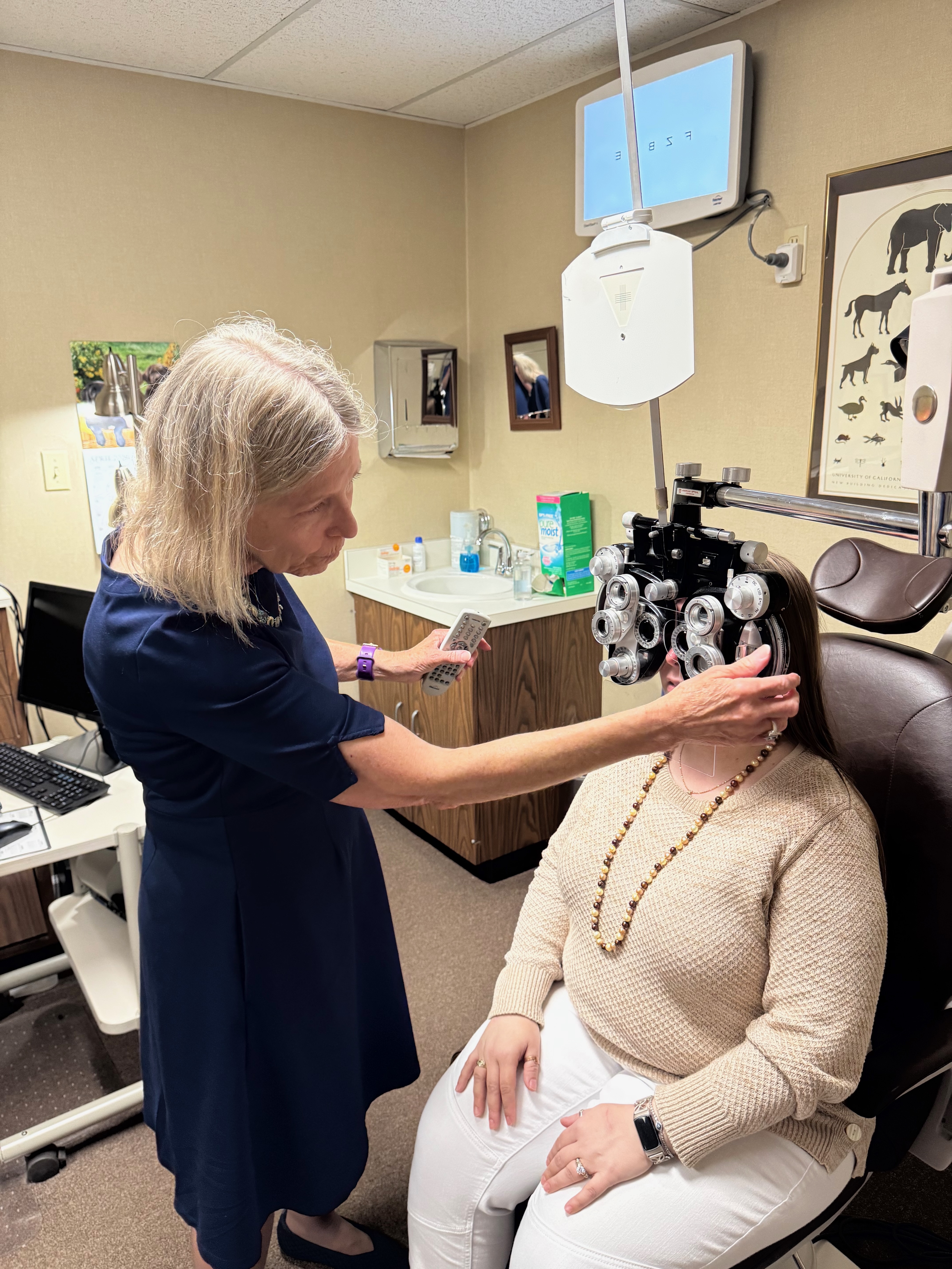
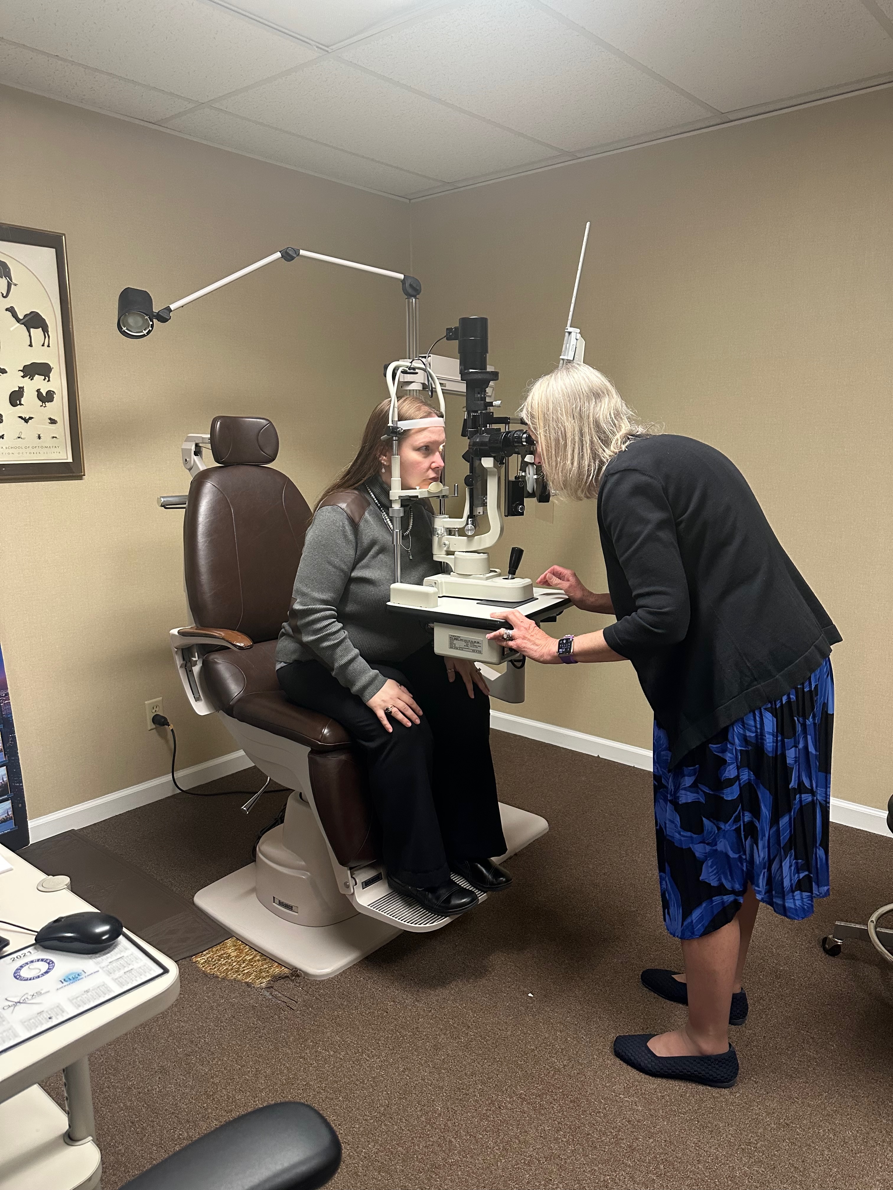
Visit 1 — the initial fitting. We do detailed topography of both eyes. We measure the cornea in several ways. We have a long conversation about your daily life — whether you work at a screen, whether you drive at night, whether you have allergies, what you've tried before. Based on all of that, I select an initial trial lens for each eye. I put the lens on, let it settle, and look at how it sits on the cornea using a slit lamp and fluorescein dye. Adjustments to the lens design are made on the spot if I can see what's needed.
Visit 2 — the trial wear. You wear the trial lenses for a week or two and come back with a report. How did they feel? How was your vision? Were there moments of fluctuation? Were they hard to insert? The trial period catches problems that don't show up in a 30-minute office visit.
Visit 3 — the dispense. Based on what we learned from the trial, we order the final custom lenses. When they arrive, we fit them carefully, teach you to insert and remove them, and send you home with a follow-up scheduled.
Visit 4 and beyond — follow-up. Specialty lenses need monitoring. We see new wearers at one week, one month, and three months in the first year, then annually after that. Any issue — discomfort, vision change, redness — and we want to see you immediately. The follow-up is not optional. It is part of the fitting.
This is why specialty fitting takes time and why it cannot be rushed. A patient who pushes for a same-day fix is a patient who ends up with a lens that doesn't fit and a bad opinion of the whole process.
What life with the lenses looks like
After the fitting period stabilizes — usually about two months in — life with specialty contact lenses is more normal than most patients expect.
Insertion takes a minute. Removal takes a few seconds. You wear them most of the day, take them out at night, clean them with the appropriate solution, and put them in a case. Scleral lenses require filling with saline before insertion, which is a small extra step. RGP and hybrid lenses do not.
You can drive with them. You can work at a computer all day with them. You can play sports with them — most sports, anyway; we have a separate conversation about contact sports and water exposure. You can go on long flights with them, though I advise patients to bring a backup pair and rewetting drops.
Most of my long-term keratoconus patients tell me they forget they have the lenses in. The vision feels normal. The eye feels normal. The keratoconus, in the day-to-day sense, becomes something they think about once a year at their annual check.
What to do next
If you have just been diagnosed with keratoconus, or if you've been told you have it but haven't seen a specialist, the next step is a thorough fitting consultation. That means topography, a full discussion of lens options, and an honest conversation about what's possible for your specific eyes.
Bring whatever records you have from your previous visits — especially any topography images, even old ones, because they tell us how your cornea has changed over time. Bring your current glasses and any contact lenses you've tried before, even if they didn't work. Bring questions written down, because you will think of more on the way home, and we can address them at the follow-up.
What you should not do is panic-search forums at 1 AM. Most of what's there is outdated, anecdotal, or worst-case. Keratoconus in 2026 is a treatable, manageable condition. The patients I fit live full lives with sharp vision. So will you.
Dr. Patricia Gelner has practiced optometry at the same Chesterfield, MO address since 1986. She trained under Dr. Paul E. Resler, one of the original Wesley Jessen contact lens fitters. Gelner Optometry specializes in custom contact lens fitting for keratoconus, post-LASIK, and other irregular corneas.
To schedule a keratoconus consultation, call 314-434-2626 or send a note.